ACL Reconstruction
Anterior Cruciate Ligament Anatomy Indications for ACL Reconstruction
The anterior cruciate ligament (ACL) is one of the major stabilising ligaments in the knee. It is a strong rope-like structure located in the centre of the knee, running from the femur to the tibia. The ACL is one of the four major ligaments of the knee that connects the femur (thighbone) to the tibia (shinbone) and helps stabilise your knee joint. It prevents excessive forward movement of the lower leg bone (tibia) in relation to the thighbone (femur) as well as limits rotational movements of the knee. When this ligament tears, unfortunately, it does not heal, and often leads to the feeling of instability in the knee. A tear in this ligament can make you feel as though your knees will not allow you to move or even hold you up.
Causes
An ACL injury most commonly occurs during sports , work or hobbies that involve twisting or overextending your knee. An ACL can be injured in several ways:
- Sudden directional change causing excessive twisting or rotation of the knee
- Slowing down while running
- Landing incorrectly from a jump
- Direct blow to the side of your knee, such as during a football tackle
- Contact injuries in collision sports or road accidents
- Hyperextension injuries
- Hyperflexion injuries
With ageing the strength of the ACL diminishes which means the cause of ACL tear may appear unremarkable.
About one in five people may be able to continue with activity following the initial injury tearing the ACL leading to an underestimation of the severity of injury.
Early Symptoms following ACL Injury
When you injure your ACL, you may hear a loud "pop" sound and feel the knee buckle meaning the knee didn’t feel stable or support their body weight. Some people have a sense of something tearing inside the knee or a sense of the knee momentarily coming apart Pain at times can be near immediate and severe although this can vary. Within a few hours and commonly up to twenty four hours after an ACL injury, your knee may swell due to bleeding from vessels within the torn ligament. Early icing may minimize swelling. You may notice that the knee feels unstable or seems to give way, especially when trying to change direction on the knee.
Late Symptoms following ACL injury
The late symptoms following ACL injury are non specific and relate to the unstable tibiofemoral joint as well as to the damage that may develop to the menisci which act as the temporary stabilisers of the knee once the ACL tears.
Altered knee biomechanics then may affect the patellar leading to anterior knee pain.
Some patients with torn ACL, intact menisci and excellent balance and neuro muscular control may initially cope well or adapt by avoidance of twisting activity .
Others have a constant sense of apprehension the knee will not support them or in some cases the knee frankly gives way leading to falls. Some people complain of a lack of trust stating “Its just not right”
Types of ACL Tear
There are three grades of ACL tear
In grade I tears , the ligament is mildly damaged and slightly stretched, but the knee joint is stable.
In grade II tears , there is a partial tear of the ligament. The ligament is in continuity but may have had an intra substance stretch or torn one of the two ACL bundles . The ACL has lost function and the knee may be unstable . Grade II tears are increasingly recognised to be a cause of symptoms following ACL injury. A ligament may undergo a complete tear but still retain continuity between disrupted fibres so it is the loss of function (resistance to displacement) that defines an ACL tear, not the property of continuity.
Partial tears may in time progress to grade III tears
In grade III tears , there is a complete tear of the ACL making the knee joint unstable. This is commonly called a rupture.
Diagnosis
ACL tears are diagnosed by the following:
- History of injury
- Nature of knee pain and other symptoms
- Physical examination
- Imaging tests
In your assessment I will:
- Take a medical history
- Perform a physical examination
- Assess the joint's range of motion
- Assess the stability of both knees .During a Lachman test, knees with a torn ACL may show an increased forward movement of the tibia and a soft or mushy endpoint compared to a healthy knee. A pivot shift test is another test that may be performed to assess an ACL tear and the rotational stability of the knee . During this test, if the ACL is torn, the tibia will move forward when the knee is completely straight and as the knee bends past 30°, the tibia shifts back into correct place in relation to the femur.
- Organise appropriate imaging tests
Imaging tests
- X-rays may be required to look for evidence of fractures or pre-existing osteoarthritis in older athletes
- MRI scans may show the ACL tear as well as any meniscal or cartilage damage.
- The accuracy of MRI is at its highest if MRI is performed within 6 weeks of initial knee injury
ACL Reconstruction Procedure
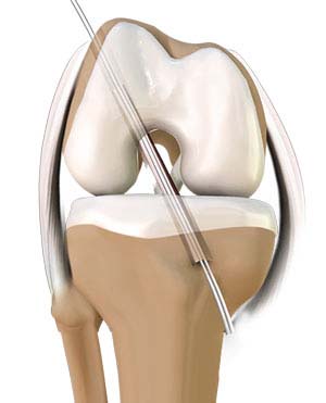
Anterior cruciate ligament reconstruction is a surgery to reconstruct the torn ligament of your knee with a tissue graft. The goal of ACL reconstruction surgery is to tighten your knee ligament and restore its stability. It is a commonly performed surgical procedure, and with recent advances in arthroscopic surgery, can be performed with minimal incisions and low complication rates.
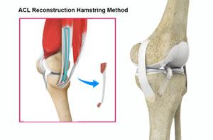
ACL reconstruction hamstring method
Anterior cruciate ligament reconstruction hamstring method is a surgical procedure to replace the torn ACL with part of the hamstring tendon taken from your own leg. The hamstring is the muscle located on the back of your thigh. The procedure is performed under general anaesthesia. Your surgeon will make small cuts, about 1/4-inch-long, around your knee. An arthroscope, a tube with a small video camera on the end, is inserted through one incision to view the inside of the knee joint. Along with the arthroscope, a sterile solution is pumped into the joint to expand it, enabling your surgeon to have a clear view and space to work inside the joint. The knee is bent at right angles and the hamstring tendons felt. A small incision is made over the hamstring tendon attachment to the tibia and the two tendons are stripped off the muscle and the graft is prepared. The torn ACL will be removed and the pathway for the new ACL is prepared. The arthroscope is reinserted into the knee joint through one of the small incisions. Holes are then drilled into the upper and lower leg bones where these bones come together at the knee joint. The holes form tunnels in your bone to accept the new ACL hamstring graft. The graft is pulled through the predrilled holes in the tibia and femur. The new tendon is then fixed into the bone with anchors to hold the ACL graft in place while the ligament heals into the bone. The incisions are then closed with sutures and a dressing is placed.
Risks and complications of ACL Reconstruction
The possible risks and complications associated with ACL reconstruction with the hamstring and patellar tendon methods include but are not limited to :
- Ongoing Instability
- Numbness of outer leg
- Infection
- Blood clots (deep vein thrombosis)
- Nerve and blood vessel damage
- Failure of the ACL graft
- Loosening of the graft
- Decreased range of motion causing knee stiffness
- Crepitus (crackling or grating feeling behind the kneecap)
- Pain in the knee
- Repeat injury to the ACL graft
- Recurrent hamstring sprains
- Instrument breakage
Postoperative care after ACL Reconstruction:
Individualised Rehabilitation
Following the surgery, rehabilitation begins immediately. A physiotherapist whom you will have met before your surgery will teach you specific exercises to strengthen your leg and restore knee movement. Avoid competitive sports or other at risk activities for 9-12 months to allow the new ACL graft to incorporate into the knee joint.
ACL Reconstruction Hamstring Method
The anterior cruciate ligament is one of the major stabilizing ligaments in the knee. It is a strong rope like structure located in the center of the knee running from the femur to the tibia. When this ligament tears unfortunately, it does not heal and often leads to the feeling of instability in the knee.
ACL reconstruction is a commonly performed surgical procedure and with recent advances in arthroscopic surgery can now be performed with minimal incision and low complication rates.
The advancements in arthroscopic surgery make it easy for surgeons to see and work on knee structures through small incisions. Repair of the torn ligament can be performed at the same time as the diagnostic arthroscopy with fewer surgical risks.
The surgery can usually be done as an outpatient procedure which means you may be discharged to go home on the same day as the procedure.
ACL Reconstruction Hamstring Tendon
Anterior cruciate ligament (ACL) reconstruction hamstring method is a surgical procedure that replaces the injured ACL with a hamstring tendon. Anterior cruciate ligament is one of the four major ligaments of the knee that connects the femur (thigh bone) to the tibia (shin bone) and helps stabilise your knee joint. Anterior cruciate ligament prevents excessive forward movement of the lower leg bone (the tibia) in relation to the thigh bone (the femur) as well as limits rotational movements of the knee.
A tear of this ligament can make you feel as though your knees will not allow you to move or even hold you up. Anterior cruciate ligament reconstruction is surgery to reconstruct the torn ligament of your knee with a tissue graft.
Causes
An ACL injury most commonly occurs during sports that involve twisting or overextending your knee. An ACL can be injured in several ways:
- Sudden directional change
- Slowing down while running
- Landing from a jump incorrectly
- Direct blow to the side of your knee, such as during a football tackle
Symptoms
When you injure your ACL, you might hear a loud "pop" sound and you may feel the knee buckle. Within a few hours after an ACL injury, your knee may swell due to bleeding from vessels within the torn ligament. You may notice that the knee feels unstable or seems to give way, especially when trying to change direction on the knee.
Diagnosis
An ACL injury can be diagnosed with a thorough physical examination of the knee and diagnostic tests such as X-rays, MRI scans and arthroscopy. X-rays may be needed to rule out any fractures. In addition, your doctor will often perform the Lachman’s test to see if the ACL is intact. During a Lachman test, knees with a torn ACL may show increased forward movement of the tibia and a soft or mushy endpoint compared to a healthy knee.
Pivot shift test is another test to assess ACL tear. During this test, if the ACL is torn, the tibia will move forward when the knee is completely straight and as the knee bends past 30° the tibia shifts back into correct place in relation to the femur.
Procedure
The goal of ACL reconstruction surgery is to tighten your knee and to restore its stability.
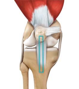
Anterior cruciate ligament reconstruction hamstring method is a surgical procedure to replace the torn ACL with part of the hamstring tendon taken from the patient’s leg. The Hamstring is the muscle located on the back of your thigh. The procedure is performed under general anesthesia. Your surgeon will make two small cuts about 1/4-inch-long around your knee. An arthroscope, a tube with a small video camera on the end is inserted through one incision to see the inside of the knee joint. Along with the arthroscope, a sterile solution is pumped into the joint to expand it enabling the surgeon to have a clear view and space to work inside the joint. The knee is bent at right angles and the hamstring tendons felt. A small incision is made over the hamstring tendon attachment to the tibia and the two tendons are stripped off the muscle and the graft is prepared. The torn ACL will be removed and the pathway for the new ACL is prepared. The arthroscope is reinserted into the knee joint through one of the small incisions. Small holes are drilled into the upper and lower leg bones where these bones come together at the knee joint. The holes’ form tunnels in your bone to accept the new graft. Then the graft is pulled through the predrilled holes in the tibia and femur. The new tendon is then fixed into the bone with screws to hold it into place while the ligament heals into the bone. The incisions are then closed with sutures and a dressing is placed.
Risks and complications
Possible risks and complications associated with ACL reconstruction with hamstring method include:
- Numbness
- Infection
- Blood clots (Deep vein thrombosis)
- Nerve and blood vessel damage
- Failure of the graft
- Loosening of the graft
- Decreased range of motion
- Crepitus (crackling or grating feeling of the kneecap)
- Pain in the knee
- Repeat injury to the graft
Post-operative care
Following the surgery, rehabilitation begins immediately. A physical therapist will teach you specific exercises to be performed to strengthen your leg and restore knee movement. Avoid competitive sports for 5 to 6 months to allow the new graft to incorporate into the knee joint.
Anterior cruciate ligament reconstruction is a very common and successful procedure. It is usually indicated in patients wishing to return to an active lifestyle especially those wishing to play sports involving running and twisting. Anterior cruciate ligament injury is a common knee ligament injury. If you have injured your ACL, surgery may be needed to regain full function of your knee.
ACL Reconstruction Patellar Tendon
Anterior cruciate ligament (ACL) reconstruction patellar tendon is a surgical procedure that replaces the injured ACL with a patellar tendon. Anterior cruciate ligament is one of the four major ligaments of the knee that connects the femur (thigh bone) to the tibia (shin bone) and helps stabilise the knee joint. Anterior cruciate ligament prevents excessive forward movement of the lower leg bone (tibia) in relation to the thigh bone (femur) as well as limits rotational movements of the knee.
A tear of this ligament can make you feel as though your knees will not allow you to move or even hold you up. Anterior cruciate ligament reconstruction is surgery to reconstruct the torn ligament of your knee with a tissue graft.
Causes
An ACL injury most commonly occurs during sports that involve twisting or overextending your knee. The ACL can be injured in several ways:
- Sudden directional change
- Slowing down while running
- Landing from a jump incorrectly
- Direct blow to the side of your knee, such as during a football tackle
Symptoms
When you injure your ACL, you might hear a loud "pop" sound and you may feel the knee buckle. Within a few hours after an ACL injury, your knee may swell due to bleeding from vessels within the torn ligament. You may notice that the knee feels unstable or seems to give way, especially when trying to change direction on the knee.
Diagnosis
An ACL injury can be diagnosed with a thorough physical examination of the knee and diagnostic tests such as X-rays, MRI scans and arthroscopy. X-rays may be needed to rule out any fractures.
In addition, your doctor will often perform the Lachman’s test to see if the ACL is intact. During a Lachman test, knees with a torn ACL may show increased forward movement of the tibia and a soft or mushy endpoint compared to a healthy knee.
Pivot shift test is another test to assess ACL tear. During the pivot shift test, if the ACL is torn the tibia will move forward when the knee is completely straight and as the knee bends past 30° the tibia shifts back into correct place in relation to the femur.
Procedure
The goal of ACL reconstruction surgery is to tighten your knee and to restore its stability.
Anterior cruciate ligament reconstruction patellar tendon is a surgical procedure to replace the torn ACL with part of the patellar tendon taken from the patient’s leg. The new ACL is harvested from the patellar tendon that connects the bottom of the kneecap (patella) to the top of the shinbone (tibia). The procedure is performed under general anesthesia. Your surgeon will make two small cuts about ¼ inch around your knee. An arthroscope, a tube with a small video camera on the end is inserted through one incision to see the inside of the knee joint. Along with the arthroscope, a sterile solution is pumped into the knee to expand it providing the surgeon a clear view of the inside of the joint. The torn ACL will be removed and the pathway for the new ACL is prepared. Your surgeon makes an incision over the patellar tendon and takes out the middle third of the patellar tendon, along with small plugs of bone where it is attached on each end. The remaining portions of the patellar tendon on either side of the graft are sutured back after its removal. Then the incision is closed. The arthroscope is reinserted into the knee joint through one of the small incisions. Small holes are drilled into the upper and lower leg bones where these bones come together at the knee joint. The holes’ form tunnels in your bone to accept the new graft. Then the graft is pulled through the predrilled holes in the tibia and femur. The new tendon is then fixed into the bone with screws to hold it into place while the ligament heals into the bone. The incisions are then closed with sutures and a dressing is placed.
Risks and complications
Possible risks and complications associated with ACL reconstruction with patellar tendon method include:
- Numbness
- Infection
- Blood clots (Deep vein thrombosis)
- Nerve and blood vessel damage
- Failure of the graft
- Loosening of the graft
- Decreased range of motion
- Crepitus (crackling or grating feeling of the kneecap)
- Pain in the knee
- Repeat injury to the graft
Post-operative care
Following the surgery rehabilitation begins immediately. A physical therapist will teach you specific exercises to strengthen your leg and restore knee movement. Avoid competitive sports for 5 to 6 months to allow the new graft to incorporate into the knee joint.
Anterior cruciate ligament reconstruction is a very common and successful procedure. It is usually indicated in patients who desire to return to an active lifestyle especially those wishing to play sports involving running and twisting. Anterior cruciate ligament injury is a common knee ligament injury. If you have injured your anterior cruciate ligament, surgery may be needed to regain full function of your knee.